


UMMC’s new 3D bioprinter brings cancer research to life
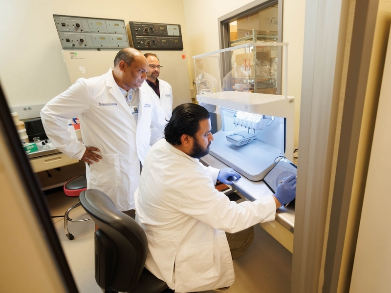
A new 3D bioprinter at the University of Mississippi Medical Center is giving cancer researchers another way to study tumors by building laboratory models that bring together the various kinds of cells found in and around them.
Equipped with six print heads, the machine can combine living cells with materials that support their growth. Unlike conventional 3D printers, which typically shape plastic or other nonliving materials, a bioprinter can create structures researchers use to examine how cells behave together.
For Dr. Jawed Siddiqui, an associate professor in UMMC's Department of Cell and Molecular Biology and a researcher with the Cancer Center and Research Institute, the technology offers a promising innovative approach to investigating cancer and potential treatments. His laboratory is helping establish bioprinting as a research tool at UMMC.
Dr. Gunjan Sharma, an instructor, and Dr. Mohd Abdullah Khan, a postdoctoral researcher, are also involved in the department’s bioprinting work. With six print heads, researchers can incorporate cancer, fat, bone and immune cells into models designed to capture aspects of a tumor’s surroundings.
Cancer cells have long been studied in flat laboratory dishes. Those experiments remain valuable, but they cannot fully reproduce the interactions among cancer cells, nearby tissues, and immune cells inside the body.
The new bioprinter gives researchers a way to study those interactions more realistically.
Sharma said researchers can use multiple print heads to incorporate different cell populations into the same model to learn how cells influence one in an environment that more closely resembles the tissues they encounter inside the body. This work aligns with major initiatives by the National Institutes of Health and guidelines promoting New Approach Methodologies, or NAMs, and human-based research to reduce animal testing.
“How does the ecosystem around a cancer cell influence what that cancer cell does?” Siddiqui asked. The answer matters because a tumor’s surroundings can affect its growth, spread and response to treatment.
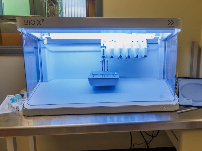
For example, a drug that kills cancer cells in a dish may work differently when those cells are surrounded by other types of tissue. More realistic laboratory models could help researchers investigate why.
That question is central to Siddiqui’s work on aggressive prostate cancer, particularly what happens when cancer spreads to bone.
His laboratory studies metastasis — the spread of cancer from its original site to another part of the body. Bone is a common site of spread for advanced prostate and breast cancers when treatment can become more difficult.
Siddiqui is investigating how cancer cells communicate with bone cells, immune cells and surrounding tissue, and how those interactions may help tumors survive or resist treatment.
His research also examines how metabolic signals, growth factors and molecular pathways help prostate cancer cells adapt to bone. By identifying vulnerabilities in those processes, the team hopes to find new therapeutic targets that could complement existing treatments.
His team also examines metabolic signals, growth factors and molecular pathways that may help prostate cancer cells adapt to bone. Finding weaknesses in those processes could point toward new treatments.
Bioprinted models could provide another way to study those interactions and test experimental therapies. Researchers would first need to establish how well the models reproduce the conditions they aim to study.
A longer-term possibility is personalized cancer research that uses cells collected from patients to build models that reflect the characteristics of their individual tumors.
That approach is not yet a proven guide to patient care. Scientists must determine whether bioprinted models accurately reflect human tumors and whether their results can reliably predict a patient’s response to treatment.
Its ability to accommodate different cell types creates opportunities for investigators in cancer biology, immunology, biomedical engineering, pharmacology, and other disciplines. The technology could also encourage collaboration among UMMC investigators in cancer biology, immunology, biomedical engineering and pharmacology.
Those experiences could help prepare the next generation of investigators to develop and apply technologies that may reshape biomedical research.
For graduate students, medical trainees and early-career scientists, the bioprinter offers hands-on experience with a developing research method.
“I think this is an important investment not just in a machine, but in the future of biomedical research at UMMC and in Mississippi,” Siddiqui said.