New technology makes it harder for cancer to hide
New technology incorporated into UMMC's Breast Imaging Services will give many women a better chance of earlier cancer detection.
The new three-dimensional software from Hologic will enable doctors to better see smaller areas of cancer in dense breast tissue.
"This is a new tool that is crucial for women with dense breasts," said Dr. Harpreet Talwar, an assistant professor of radiology and chief of UMMC's breast imaging division. "Because of the volume of breast tissue, cancer can hide from two-dimensional images. It has less chance of hiding on the 3-D images."
Currently most women receive 2-D imaging.
"When a 2-D image isn't clear enough for a radiologist to say with certainty that it depicts no cancer, the woman may be recalled to have additional imaging or sonogram," Talwar said. "In dense breast tissue, 3-D can detect cancer with more clarity and confidence. With 2-D images, the same cancer may be harder to detect and can be missed."
Willie Smith was among the first to try it at UMMC. A colon cancer and sickle cell survivor, the Clinton resident said she believes in getting the best screening possible.
"They said you could see more with 3-D," she said of Talwar and the breast imaging staff. "I said I want to go for it."
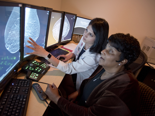
Recently, she sat with Talwar and listened as the radiologist explained her images.
"Think of it like a book," Talwar said. "Before we could see the front and back cover. The pages in the middle were projected on top of each other. Now we can flip through and see the pages of the book."
"You really can see more," Smith said, watching as Talwar compared Smith's 2-D and 3-D images.
Regular 2-D mammograms for women with dense tissue may be harder to interpret, so 3-D images often can allow radiologists to distinguish between malignant and non-malignant shapes, Talwar said.
"We're figuring out what lies inside the breast instead of looking at a compressed 2-D picture," she said. "The recall rates with tomosynthesis or 3-D examination are significantly lower than with 2-D imaging because the confidence of interpretation and cancer detection using 3-D technology is higher compared to 2-D."
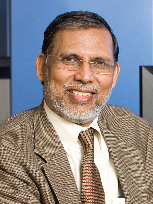
"This is a great example of how medical advances are improving the health of Mississippians by using technology that improves detection rates of patients with cancer and more accurately excludes patients that don't have the disease," said Dr. Timothy McCowan, Department of Radiology chair.
It's one more step toward improving services for patients, said Dr. Srinivasan Vijayakumar, Cancer Institute director.
"Our goal is to always find better ways to detect cancer earlier," said Vijayakumar, also Department of Radiation Oncology chair. "And on the other side, in many cases, this technology can relieve the stress of being called back for more imaging."
The American Journal of Roentgenology published a study that showed the 3-D mammograms resulted in:
- A drop in the recall rate. Women who had no cancer did not have to return for more imaging just because the 2-D imaging was harder to interpret.
- A drop in biopsy rates. The 3-D imaging more clearly showed no cancer was present.
- An increase in cancer detected.
- An increase in invasive cancer detected.
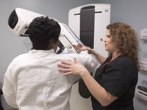
So how does this work? Talwar said a woman has a traditional 2-D mammogram, then the same machine rotates around her breast in an arc, taking images at predetermined intervals. A computer combines the images. Then, radiologists can look at them in much thinner sections.
While the 3-D imaging has slightly higher radiation doses than traditional 2-D imaging, it is still under the FDA limit for mammography.
Radiologists and technicians have completed training and working with the new technology, Talwar said.
In Mississippi, Medicare and many insurance providers will pay for the 3-D imaging. Women who need this type of imaging should check with their insurers to see if it is covered, Talwar said.