Breast cancer early detection, treatment saves lives
You’ve discovered a lump in your breast. What do you do next?
Don’t panic, but instead make a plan to get it checked, University of Mississippi Medical Center providers say.

“The next step would be to contact your primary care provider or gynecologist,” said Dr. Scott Berry, chief of breast surgery at the University of Mississippi Medical Center and surgical director of UMMC’s Comprehensive Breast Center.
Finding a lump is a scary thing, but it’s not a time to panic, Berry said. “Get in to be evaluated,” he said.
That will entail giving details to the provider about the lump or mass and talking about next steps, which usually include a mammogram or ultrasound, he said. “If there’s something there, the important thing to know is that most lumps in the breast are not cancer,” Berry said.
UMMC’s Cancer Center and Research Institute uses the latest diagnostic techniques to detect and treat cancer as early and accurately as possible, including diagnostic mammogram, 3-D mammogram, ultrasound and magnetic resonance imaging.
In fact, Berry said, an important part of treatment is to give women who don’t have cancer peace of mind. “When we see someone, we can get a mammogram and studies done within days,” Berry said. “You may be able to answer the questions right then.”
If a screening shows an abnormality or something concerning, a number of things can happen next:
- A radiologist will look at its shape and layout to judge if it’s cancerous or not by using diagnostic imaging such as an ultrasound, mammogram or a breast MRI.
- If the abnormality is still a concern, a biopsy is usually required. Depending on the recommendation of providers, it could be a stereotactic needle-guided biopsy in which the patient lies face down on a table with the breast suspended through a hole and compressed. Digital mammography is used to locate the mass in the breast, and then the doctor uses a small needle to remove tissue samples.
It could be a surgical biopsy, which is rare. Sometimes the surgeon will use an incisional biopsy, which removes part of the mass, or an excisional biopsy, which removes the entire mass. A pathologist will then examine the tissue.
A patient could have a tomosynthesis (3-D) guided biopsy, a minimally invasive procedure using X-ray images to gather tissue samples from the breast. The physician is able to locate and target specific areas of breast tissue for investigation.
Or, it could be an ultrasound-guided needle biopsy in which doctors use ultrasound to help them guide a needle into the right spot to collect a tissue sample.
- If cancer is detected, a CT scan, PET scan, PET/CT scan, or bone scan may be used to see if the cancer has spread.
If you’re told you have or may have breast cancer, the American Cancer Society recommends you ask these questions: Why do you think I have cancer? Is there a chance I don’t have cancer? Would you write down the kind of cancer you think I might have? What will happen next?
Many patients choose to seek a second option on a cancer diagnosis, something UMMC physicians do by reviewing diagnostic images sent from other imaging locations outside the Medical Center.
Women should perform self-examinations, even if they show no signs or symptoms. Do a self-exam every month, just before your period, as recommended by the American College of Obstetrics and Gynecology. Physicians at the UMMC Cancer Center and Research Institute recommend regular breast exams beginning in young adulthood and annual mammograms beginning at age 40.
The female breast cancer death rate has declined by 42% as of 2019, mainly because of earlier detection and improved treatment, the American Cancer Society says.

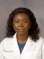
Women who find a lump should not immediately assume the worst, but should see their provider promptly, said Dr. Carmen Obeng, a resident in the Department of Preventive Medicine at the John D. Bower School of Population Health.
Those under age 30 usually have more dense breasts and often undergo ultrasound, Obeng said. “If it shows there is something there, you will usually have a follow-up mammogram,” she said.
Early detection, she said, is key to success in treating breast cancer, and so is knowing your genetic history of the disease and predisposition to having it. The most common cause of hereditary breast cancer is an inherited mutation in the BRCA1 or BRCA2 gene.
“If you are at high risk because of a family history of breast cancer, this is a conversation you need to have with your provider,” Obeng said. Early mammograms are recommended for those at high risk, regardless of age, she said.
Patients with a close family history of breast cancer can talk to a UMMC medical geneticists who performs hereditary cancer syndrome counseling and pre-symptomatic testing for breast cancer.
For more information about breast cancer care at UMMC, click here. To make an appointment, call (888) 815-2005. To schedule a screening, call (601) 815-4723.
The above article appears in CONSULT, UMMC’s monthly e-newsletter sharing news about cutting-edge clinical and health science education advances and innovative biomedical research at the Medical Center and giving you tips and suggestions on how you and the people you love can live a healthier life. Click here and enter your email address to receive CONSULT free of charge. You may cancel at any time.


