|
Dr. Joey Granger, Billy S. Guyton Distinguished Professor of physiology and biophysics, is the 2016 Southeastern Conference Faculty Achievement Award winner for the University of Mississippi.
|

|
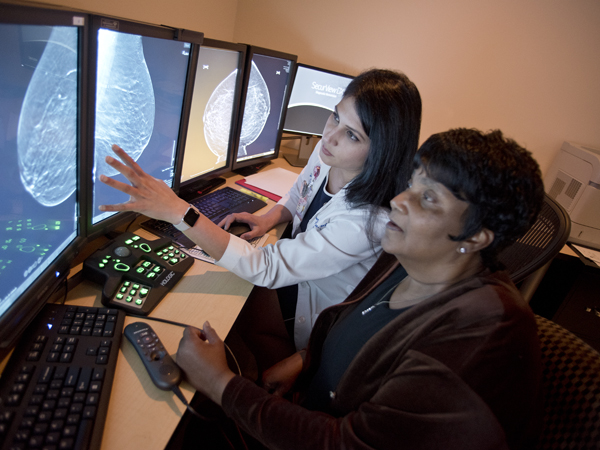
New technology incorporated into UMMC's Breast Imaging Services will give many women a better chance of earlier cancer detection.
|
|
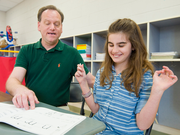
“As long as you are in school, your job is to make good grades.” This is a phrase heard from the mouth of many a parent. A kid's occupation is to learn, play and grow.
|

|

|
The Medical Center is proud to announce the following additions to its faculty and leadership staff:
|
|
The Society for Academic Emergency Medicine has selected not one, but two Medical Center faculty for prestigious national awards that highlight their contributions and commitment to emergency medicine.
|

|


